Cell imaging of the liver may go beyond still pictures and evolve into real-time videos in the future, providing doctors continuous and dependable information on the inner workings of their patients and possibly directions on how to cure them, the National Science Council (NSC) said yesterday.
The technology is called in vivo multiphoton microscopy and has achieved laboratory success. A team of National Taiwan University medical and physics researchers installed a titanium intravital hepatic (liver) imaging chamber on the belly of a rat and was able to video monitor metabolic activities of its liver.
The team's work was published in the Journal of Biomedical Optics last in February last year.
"Multiphoton microscopy is a relatively new nonlinear optical technique developed in the early 19 90s and allows for effective label-free imaging of many tissue types," said Wesley Chen (
live action
"The technology is exciting as before we could only infer what happened inside the liver based on images taken at different times, but now we can see it in action," said Lee Hsuan-shu (李宣書), the team's contributing medical researcher.
The window-like device sheds light on liver function mechanisms, pathology and molecular biology, Lee said.
For example, the team has seen liver damage caused by Panadol overdose and has documented through bile duct ligation that bile duct congestion could cause jaundice.
"When the duct is congested, we can see that bile elimination is blocked; the juices then flow into the blood, and the reddish bilirubin and yellowish biliflavin show through the skin," Lee said.
Current imaging techniques, such as ultrasound, CT scans and magnetic resonance imaging, only provide macro images, Lee said, adding that although intravital videomicroscopy could provide images at a more microscopic level, it could not zoom in on a single cell.
"When observing at the cell level, resolution is often more important than magnification," Chen said.
less invasive
In addition to offering clearer images, the technology utilizes photons with long wavelengths and is therefore less invasive and more in-depth, Chen said.
"Compared to a single photon microscopy, which excites fluorophores with one single short-wavelength photon, multiphoton microscopy excite fluorophores with the energy of two long wavelength photons that arrive simultaneously," Chen said.
Asked when the technique could begin clinical trial, Lee said that installing a "window" in a person's abdomen might not get the greenlight from the hospital's ethics committee.
However, the "technology is promising and could be adapted in endoscopes to provide valuable information on patient diagnosis and treatment," Lee said.
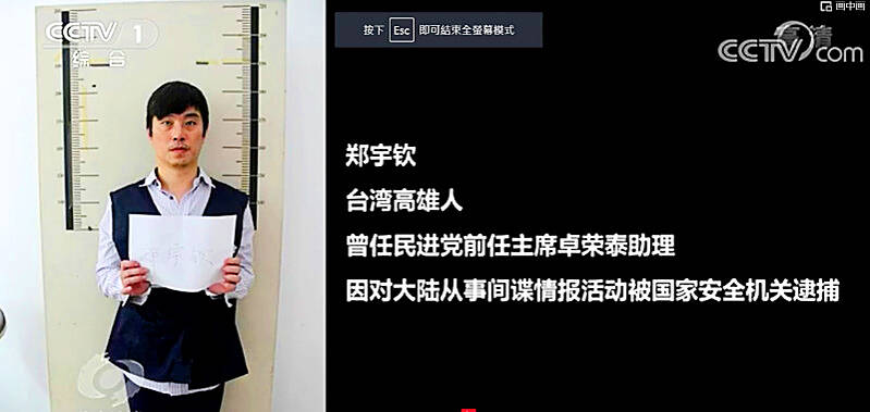
Former Czech Republic-based Taiwanese researcher Cheng Yu-chin (鄭宇欽) has been sentenced to seven years in prison on espionage-related charges, China’s Ministry of State Security announced yesterday. China said Cheng was a spy for Taiwan who “masqueraded as a professor” and that he was previously an assistant to former Cabinet secretary-general Cho Jung-tai (卓榮泰). President-elect William Lai (賴清德) on Wednesday last week announced Cho would be his premier when Lai is inaugurated next month. Today is China’s “National Security Education Day.” The Chinese ministry yesterday released a video online showing arrests over the past 10 years of people alleged to be
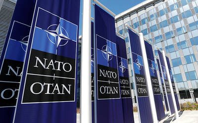
THE HAWAII FACTOR: While a 1965 opinion said an attack on Hawaii would not trigger Article 5, the text of the treaty suggests the state is covered, the report says NATO could be drawn into a conflict in the Taiwan Strait if Chinese forces attacked the US mainland or Hawaii, a NATO Defense College report published on Monday says. The report, written by James Lee, an assistant research fellow at Academia Sinica’s Institute of European and American Studies, states that under certain conditions a Taiwan contingency could trigger Article 5 of NATO, under which an attack against any member of the alliance is considered an attack against all members, necessitating a response. Article 6 of the North Atlantic Treaty specifies that an armed attack in the territory of any member in Europe,
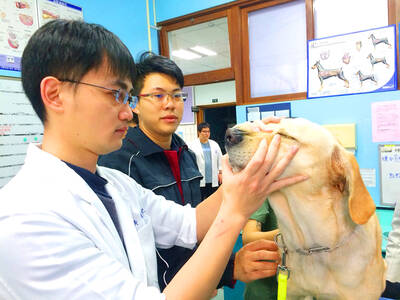
LIKE FAMILY: People now treat dogs and cats as family members. They receive the same medical treatments and tests as humans do, a veterinary association official said The number of pet dogs and cats in Taiwan has officially outnumbered the number of human newborns last year, data from the Ministry of Agriculture’s pet registration information system showed. As of last year, Taiwan had 94,544 registered pet dogs and 137,652 pet cats, the data showed. By contrast, 135,571 babies were born last year. Demand for medical care for pet animals has also risen. As of Feb. 29, there were 5,773 veterinarians in Taiwan, 3,993 of whom were for pet animals, statistics from the Animal and Plant Health Inspection Agency showed. In 2022, the nation had 3,077 pediatricians. As of last

XINJIANG: Officials are conducting a report into amending an existing law or to enact a special law to prohibit goods using forced labor Taiwan is mulling an amendment prohibiting the importation of goods using forced labor, similar to the Uyghur Forced Labor Prevention Act (UFLPA) passed by the US Congress in 2021 that imposed limits on goods produced using forced labor in China’s Xinjiang region. A government official who wished to remain anonymous said yesterday that as the US customs law explicitly prohibits the importation of goods made using forced labor, in 2021 it passed the specialized UFLPA to limit the importation of cotton and other goods from China’s Xinjiang Uyghur region. Taiwan does not have the legal basis to prohibit the importation of goods